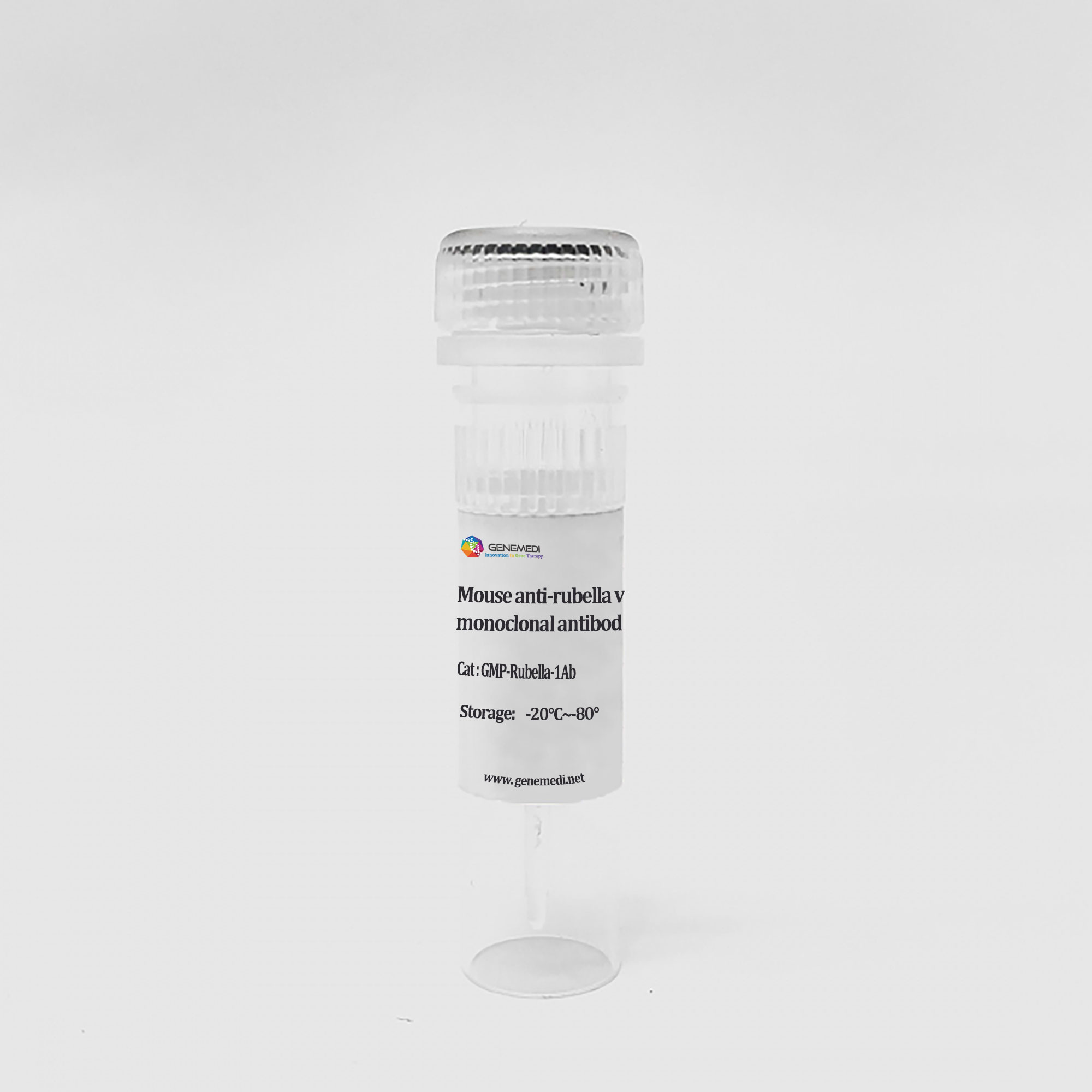
AAV9 vector system (AAV9 expression system, AAV9 packaging plasmid system )
$2,028.00
GeneMedi’s AAV9 Vector System (AAV serotype 9 helper-free packaging plasmids system) is including AAV9 Rep-Cap plamid (AAV9-RC plasmid, or called AAV-RC9 plasmid), AAV helper plasmid and AAV expression vectors (overexpression or shRNA).
GeneMedi’s AAV expression vectors have been inserted with differernt expression cassettes, containing kinds of verified protomters and reporters including GFP, zsgreen, RFP, mcherry and luciferase. The GeneMedi’s AAV expression vectors have been proved very suitalble for unique gene overexpression or shRNA-mediated knock-down (also called RNAi (RNA interference ). You can also achieve gene knock-out(KO) or gene editing using our Crispr-cas9-gRNA AAV expression vector.
AAV9 Rep-Cap plamid supplies the AAV2 Rep(replication) proteins and the AAV9 capsid protein.
The tissue tropism of AAV9 vector has been validated in neuron (CNS), lung, liver, heart and muscle, with potential applications in tissue-specific gene therapy.
AAV9 vector tissue tropism and gene transduction (serotype-specific AAV infection)
The tissue tropism of AAV9 vector has been validated in neuron(CNS), heart, muscle, retina and Pancreas, with potential applications in tissue-specific gene therapy.
.webp)
Virus and titer: AAV9-GFP, AAV9-pTBG-Luciferase (AAV-Luc), 1.4×10¹² vg/ml
Animal: mouse, C57, 2 months
Gene delivery method: tail vein, 100μl
Determine assay: 3 weeks post infection, frozen section, immunofluorescence microscopy, in vivo imaging
Conclusion: intravenous injection of AAV9-pTBG-Luciferase (AAV-Luc) only infects liver cells and no non-specific infection

Virus and titer: AAV9-GFP, 1×10¹² vg/ml
Animal: Rat, SD, 2 months (Figure 8A, B, C); mouse, C57, 8 months (Figure 8D)
Infection site: heart
Gene delivery method: myocardial in situ injection, 10μl/site, 5 sites in total (Figure 8A, B, C);intraorbital intravenous injection (Figure 8D)
Determine assay: 3 weeks post infection, frozen section, immunofluorescence microscopy

Virus and titer: AAV9-GFP, 1×10¹² vg/ml
Animal: Mouse, C57, 2 months
Infection site: Brain
Gene delivery method: Brain localization injection, 1μl
Determine assay: 3 weeks post infection, frozen section, immunofluorescence microscopy

Virus and titer: AAV9-GFAP-GFP, 1×10¹² vg/ml
Animal: mouse, C57, 2 months
Infection site: Hippocampus
Gene delivery method: Brain localization injection, 1μl
Determine assay: 3 weeks post infection, wholemount, immunofluorescence microscopy
-and-AAV9-GFP.webp)
Virus and titer: AAV9-Luciferase (AAV-Luc), AAV9-GFP, 1×10¹² vg/ml
Animal: Mouse, C57, 2 months
Infection site: Skeletal muscles
Gene delivery method: Muscle in situ injection, 10μl/site, 4 sites in total
Determine assay: 4 weeks post infection, in vivo imaging, frozen section, immunofluorescence microscopy

Virus and titer: AAV-DJ-GFP, AAV9-GFP, 1×10¹² vg/ml
Animal: mouse, C57, 2 months
Infection site: Kidney
Gene delivery method: Multiple sites injection in kidney 10μl/site, 6 sites in total
Determine assay: 3 weeks post infection, frozen section, immunofluorescence microscopy

Virus and titer: AAV2 (left) /AAV-DJ (middle) / AAV9 (right), 1×10¹² vg/ml
Animal: Nude mouse, 2 months
Infection site: Subcutaneous transplant of bowel cancer cells
Gene delivery method: Tumor injection, 10μl/site, 4 sites in total
Determine assay: 3 weeks post infection, frozen section, immunofluorescence microscopy

Virus and titer: AAV9-GFP, 1×10¹² vg/ml
Animal: mouse, C57, 2 months
Infection site: Mammary fat pad
Gene delivery method: Breast injection, 10μl/site, 4 sites in total
Determine assay: 3 weeks post infection, frozen section, immunofluorescence microscopy

Virus and titer: AAV2 (left) /AAV-DJ (middle) / AAV9 (right), 1×10¹² vg/ml
Animal: Nude mouse, 2 months
Infection site: Subcutaneous transplant of bowel cancer cells
Gene delivery method: Tumor injection, 10μl/site, 4 sites in total
Determine assay: 3 weeks post infection, frozen section, immunofluorescence microscopy

Virus and titer: AAV1/AAV6/AAV8/AAV-Rh10/AAV-DJ/AAV9-GFP, 1×10¹² vg/ml
Cells: HEK-293T
MOI: MOI=1×10⁴
Determine assay: 36 hours post infection, immunofluorescence microscopy
| Product Name | AAV9 vector system |
|---|---|
| Cat.No. |
Related products
AAV rap-cap plasmids
AAV rap-cap plasmids
AAV1 Rep-Cap plasmid (serotype 1- specific AAV RC1 plasmids)
AAV rap-cap plasmids
AAV2 variant (Y444F) Rep-Cap plasmid (retina-validated serotype AAV-RC plasmid)
AAV rap-cap plasmids
AAV-DJ/8 Rep-Cap plasmid (serotype DJ/8-specific AAV RC-DJ/8 plasmids)


